
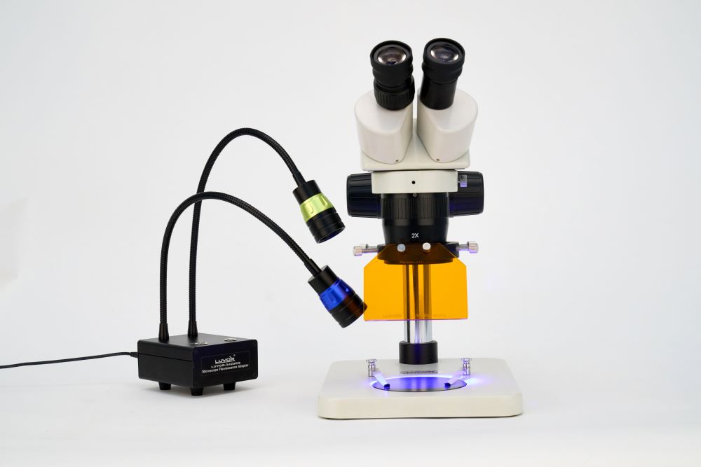
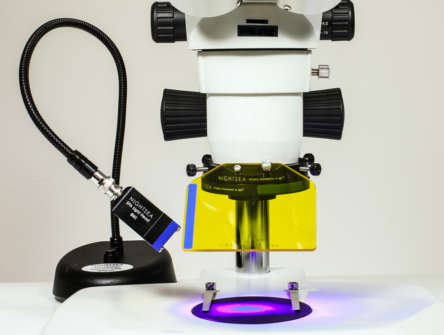
荧光适配器用于样品的预筛选
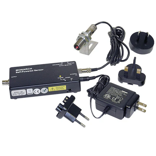
美国NIGHTSEA SFA-RB体视显微镜荧光适配器可以将您的常规实验室体视显微镜变成一种有价值的工具,用于在进入更高分辨率的荧光显微镜或共聚焦显微镜之前预先筛选样品制备的荧光。上海峰志仪器有限公司代理销售美国nightsea体视显微镜荧光适配器和荧光手电筒。有关体视显微镜荧光适配器产品介绍请浏览《美国nightsea荧光适配器 》,上海峰志仪器有限公司销售的荧光观察设备能够观察植物愈伤、叶片、种子、根系等上面的转基因的表达,还能观察转基因在动物如老鼠、斑马鱼、果蝇等实验动物上的表达。有便携式单波长荧光手电筒,也有手持式大面积照射高强度的双波长激发光源,上海峰志提供免费试机,有兴趣的老师和同学可按照网页底部联系联系。
挑战
生物样品的高分辨率成像主要基于荧光技术。共聚焦、双光子和高分辨率复合荧光显微镜几乎总是有限的资源。它们通常仅位于成像核心设施中,并且可按计划按使用付费。将荧光团引入样品的过程并不总是成功的。染色,将含GFP的质粒引入细胞,免疫组织化学 - 这些都是错误的。花时间在高端系统上寻找荧光并不罕见,而那里甚至没有发现任何荧光。
实用的解决方案
美国NIGHTSEA SFA-RB可以在标准体视显微镜上对标本进行荧光预筛选。您看到的细节并不重要 - 荧光的简单存在或不存在以及一般位置可以让您知道是否值得将标本带到成像核心。在使用费的直接费用和浪费在查看非荧光样品的时间之间,NIGHTSEA系统不需要很多节省的旅行来支付自己。
一位研究人员的工作要求用Alexa Fluor 488 Phalloidin染色兔子腰大肌纤维。对于没有沾染污渍的样品,有些令人沮丧。在获得SFA-RB荧光适配器后,她写道:
"利用NIGHTSEA SFA-RB体视显微镜荧光适配器预筛选是一种很好的方法,可以在我们尝试在共聚焦上以更高的放大倍率对肌肉纤维进行成像之前快速检查染色是否成功。

兔腰大肌纤维在白光和荧光下用Alexa Fluor 488 Phalloidin染色。使用NIGHTSEA荧光适配器的白光LED(左)和皇家蓝激发(SFA-RB)/发射光+滤光片组制作的图像。样本由布朗大学的Beth Brainerd博士和Natividad Chen博士提供。
另一位研究人员使用斑马鱼作为一个系统来研究不同的有毒物质(药物,农药,食品添加剂等)如何改变大脑发育。他写道:
"在使用NIGHTSEA SFA-RB荧光适配器筛选样品之前,我必须选择要安装的样品,去共聚焦,然后希望我的一些样品实际上是荧光的。现在我使用NIGHTSEA体视显微镜荧光适配器来预筛选我的样品,通过确保我成像的样品是荧光的,我节省了时间和金钱。

转基因斑马鱼大脑共聚焦图像
转基因斑马鱼(Dania rerio)大脑的共聚焦图像。Kaede蛋白 – 绿色是未转换的,红色是光转换的。图片由布朗大学Creton Lab的Robert Thorn提供。
英文原文:
Pre-Screening Samples for Fluorescence
The NIGHTSEA Model SFA Stereo Microscope Fluorescence Adapter can turn your routine laboratory stereo microscope into a valuable tool for pre-screening your sample preparations for fluorescence before moving on to higher resolution systems.
The Challenge
High resolution imaging of biological samples is heavily based on fluorescence techniques. Confocal, 2-photon, and high resolution compound fluorescence microscopes are almost always a limited resource. They are often located only in imaging core facilities and accessible on a scheduled, pay-per-use basis.
The processes for introducing fluorophores to specimens are not always successful. Staining, introduction of GFP-bearing plasmids to cells, immunohistochemistry – all are fallible. It is not unusual to spend time searching for fluorescence on a high end system when there is not even any there to be found.
The Practical Solution
The NIGHTSEA SFA enables fluorescence pre-screening of specimens on a standard stereo microscope. The detail that you see is not important – the simple presence or absence and general location of fluorescence lets you know whether it is worth taking your specimen to the imaging core. Between the direct expense of the use fee and the time wasted to look at a non-fluorescent specimen it will not take many saved trips for the NIGHTSEA system to more than pay for itself.
One researcher’s work requires staining rabbit psoas muscle fibers with Alexa Fluor 488 Phalloidin. There was some frustration with samples that did not take up the stain. After acquiring the SFA she wrote:
“The NIGHTSEA fluorescence setup is a great way to quickly check whether the stain was successful before we try to image the muscle fiber at a higher magnification on the confocal.”
Rabbit psoas muscle fibers stained with Alexa Fluor 488 Phalloidin
Rabbit psoas muscle fibers stained with Alexa Fluor 488 Phalloidin, in white light and fluorescence. Images made using NIGHTSEA’s white LED (left) and the Royal Blue excitation/emission light+filter set. Samples courtesy of Dr. Beth Brainerd and Natividad Chen, Brown University.
Another researcher uses zebrafish as a system to look at the way different toxicants (pharmaceuticals, pesticides, food additives, etc.) alter brain development. He writes:
“Before using NIGHTSEA to screen my samples, I would have to select samples to mount, go to the confocal and then hope that some of my samples were actually fluorescent. Now that I use NIGHTSEA to prescreen my samples I save both time and money by making sure the only samples I image are fluorescent.”
Confocal image of brain of transgenic zebrafish
Confocal image of brain of transgenic zebrafish (Dania rerio). Kaede protein – green is unconverted, red is photoconverted. Image courtesy of Robert Thorn, Creton Lab, Brown University.

 添加微信咨询!