


使用 calcofluor white钙荧光白和手持式紫外线手电筒快速检测真菌
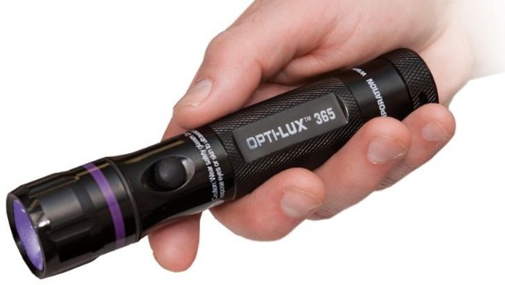
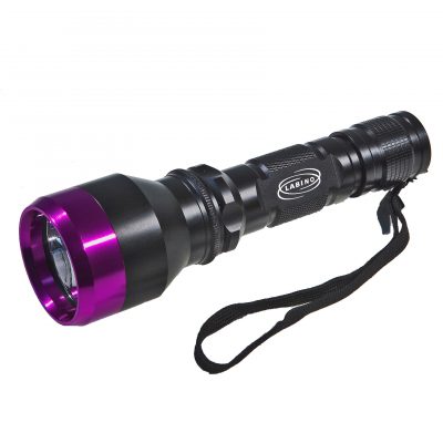
氢氧化钾 (KOH) 制剂是一种广泛用于诊断皮肤癣菌感染的方法。然而,根据用户体验和性能,KOH 具有广泛变化的敏感性和特异性。Calcofluor white (CFW) 是一种荧光蓝染料,常用于微生物实验室进行真菌鉴定。它与真菌细胞壁中的几丁质结合,并在暴露于紫外线 (UV) 光时发出明亮的荧光(图 1)。尽管被认为是皮肤癣菌检测的一种优越方法,但该技术的床边使用受到与荧光显微镜相关的成本和复杂性增加的限制。我们的研究提出了一种使用改良版 KOH 显微镜在床边识别皮肤癣菌感染的新方法:使用荧光化合物 CFW 和廉价且易于获得的手持式 395nm 紫外线手电筒作为一种有效的检测手段。

图 1用 calcofluor white 染色并用手持紫外线手电筒照射时出现阳性皮肤刮擦外观的代表性图像。注意与背景荧光相比,明亮的荧光菌丝。
这项研究得到了约翰霍普金斯医学机构审查委员会的批准。在对临床上明显的皮肤癣菌感染和对照进行皮肤刮片后,通过添加 1% CFW 溶液 (Sigma-Aldrich) 或 20% KOH 制备样品并静置 3 至 5 分钟。以传统方式检查样品,然后关闭舞台灯并使用手持式紫外光源进行切向照明(图 2)。为了比较这些方法,拍摄了一系列 30 张图像,显示以下内容:5 个阳性 KOH 制剂,5 个阴性 KOH 制剂,5 个阳性 calcofluor 制剂,具有 4',6-diamidino-2-phenylindole (DAPI) 荧光,5 个阴性 calcofluor 制剂使用 DAPI 荧光,使用手持式紫外线手电筒进行 5 次阳性 calcofluor 制剂,使用手持式紫外线手电筒进行 5 次阴性 calcofluor 制剂。DAPI 照明传统上与 CFW 一起使用,因此被用作紫外线手电筒的控制。这些图像被随机化并纳入在线 Qualtrics 调查,并分发给约翰霍普金斯大学皮肤科的主治医师和住院医师。

图 2使用手持紫外线手电筒对 calcofluor white 染色载玻片进行切向照明的示意图。
在完成调查的 20 人中,40% 是住院医师,60% 是主治医师。我们的队列在 90.5% 的时间内正确分类了 KOH 制剂。相比之下,用 DAPI 荧光照射的 CFW 和用紫外手电筒照射的 CFW 的正确分类率为 99%。此外,我们的队列确定使用 CFW 与 KOH 的图像对于真菌元素的图像是阳性还是阴性显着更快,无论照明方法如何(平均时间 7.7 秒对 16.8 秒,P < .05,配对样本t检验)。
与传统的 KOH 显微镜相比,我们的研究确定了一种使用 CFW 鉴定床边皮肤癣菌的更有效和准确的方法。尽管应限制对眼睛的照射,但使用LED紫外线手电筒(通常在 395 nm 范围内)不太可能导致严重的眼部损伤。3临床医生可以使用紫外线安全护目镜来降低这种风险,这种护目镜(紫外线防护)可以滤除 <400 nm 的紫外光。尽管这项研究受到其小规模的限制,但已经启动了一项全国性的后续研究,以评估这种检测技术的有效性和使用情况,并将我们的方法与使用其他可能增强 KOH 显微术的染料进行比较。
以上中文内容由谷歌翻译软件翻译,文章原文:
Rapid detection of fungal elements using calcofluor white and handheld ultraviolet illumination
To the Editor: Potassium hydroxide (KOH) preparation is a widely used method for diagnosing cutaneous dermatophyte infections. However, KOH has widely variable sensitivity and specificity based on user experience and performance.1 Calcofluor white (CFW) is a fluorescent blue dye commonly used in microbiology laboratories for fungal identification. It binds to the chitin in the fungal cell wall and brightly fluoresces on exposure to ultraviolet (UV) light (Fig 1). Although considered a superior method for dermatophyte detection, bedside use of this technique is limited by the increased cost and complexity associated with fluorescence microscopes.2 Our study presents a new method for bedside identification of dermatophyte infections by using a modified version of KOH microscopy: using the fluorescent compound CFW and an inexpensive and easily available handheld 395-nm light-emitting diode flashlight (TaoTronics model TT-FL001, TaoTronics, Sunvalley Group, Shenzhen, China) as an effective means of detection.
Figure thumbnail gr1
Fig 1A representative image of the appearance of a positive skin scraping when stained with calcofluor white and illuminated with a handheld ultraviolet flashlight. Note the brightly fluorescent hyphae compared with the background fluorescence.
View Large ImageFigure ViewerDownload Hi-res imageDownload (PPT)
This study was approved by the Johns Hopkins Medicine institutional review board. After performing skin scrapings of a clinically apparent dermatophyte infection and a control, samples were prepared by addition of either 1% CFW solution (Sigma-Aldrich) or 20% KOH and allowed to sit for 3 to 5 minutes. The samples were examined in the traditional manner and then with stage-light off and tangential illumination from a handheld UV light source (Fig 2). To compare these methods, a series of 30 images were captured displaying the following: 5 positive KOH preparations, 5 negative KOH preparations, 5 positive calcofluor preparations with 4′,6-diamidino-2-phenylindole (DAPI) fluorescence, 5 negative calcofluor preparations with DAPI fluorescence, 5 positive calcofluor preparations with a handheld UV flashlight, and 5 negative calcofluor preparations with a handheld UV flashlight. DAPI illumination is traditionally used with CFW and was therefore used as a control for the UV flashlight. These images were randomized and incorporated into an online Qualtrics survey and distributed to attending physicians and residents at the Johns Hopkins Department of Dermatology.
Figure thumbnail gr2
Fig 2A schematic representation showing tangential illumination of a calcofluor white–stained slide with a handheld ultraviolet flashlight.
(Courtesy of Leon Denny.)
View Large ImageFigure ViewerDownload Hi-res imageDownload (PPT)
Of the 20 individuals who completed the survey, 40% were resident physicians and 60% attending physicians. Our cohort correctly classified the KOH preparation 90.5% of the time. In contrast, the CFW illuminated with DAPI fluorescence and the CFW illuminated with the UV flashlight were both correctly classified 99% of the time. Additionally, our cohort determined if the images were positive or negative for fungal elements significantly faster for images using CFW versus KOH, regardless of the method of illuminated (mean time 7.7 seconds vs 16.8 seconds, P < .05, paired samples t test).
Our study identifies a more efficient and accurate method for bedside cutaneous dermatophyte identification using CFW compared with that of traditional KOH microscopy. Although exposure to the eyes should be limited, the use of a UV light-emitting diode flashlight, which is typically in the 395-nm range, is unlikely to result in significant ocular damage.3 Clinicians can mitigate such risk with the use of UV safety goggles, which filter out light <400 nm. Although this study is limited by its small size, a nationwide follow-up study has been initiated to assess the effectiveness and use of this detection technique and to compare our approach with the use of other dyes that may enhance KOH microscopy.
推荐浏览更多荧光蛋白表达的效果图:(荧光蛋白表达的案例展示)

 添加微信咨询!